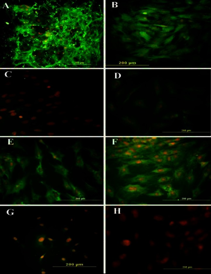
Fig. 1.
Immunocytochemistry representation of different cell markers. Following the treatment of bone marrow stromal cells with DMSO-retinoic acid at pre-induction stage, the cells were labeled with primary antibodies, followed by incubation of FITC-conjugated secondary antibody and counterstaining with ethidium bromide. Immunostained cells with (A) anti-fibronectin antibody, (B) anti-CD90 antibody, (C) anti-CD45 antibody, (D) anti-CD106 antibody, (E) anti-nestin, (F) anti-neurofilament 160 (NF-160) antibody, (G) anti-NF68 antibody and (H) anti-glial fibrilliary acidic protein antibody

