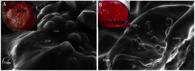
Figure 2.
Scanning electron micrograph images of alginate-gelatin (AG) cryogel scaffolds showed visible extra cellular matrix (ECM) deposition during chondrocyte culture. Image A shows cells embedded within the ECM secreted by chondrocytes over the period of 30 days on the AG scaffolds with α-KG (inset digital image of ECM deposition). Image B (control) AG cryogel scaffolds without α-KG (inset digital image of the control).

