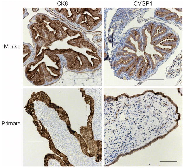
Figure 1.
Comparison of the location of FTE cells in primates and rodents. Distal fallopian tube fimbriae (primates) or oviduct (mouse) were analyzed by immunohistochemistry for an epithelial protein (CK8) or a tubal specific protein (OVGP1). In primates, FTE are on the outer surface of the stromal component of the tissue, and in mice, the FTE line the lumen of the oviductal tube.

