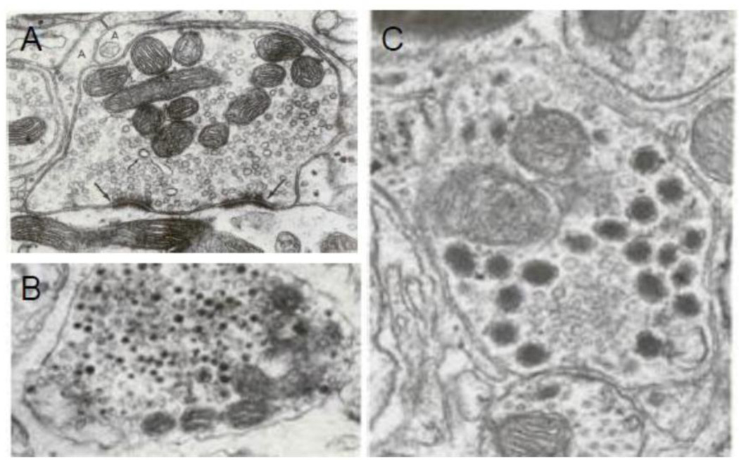
Figure 3.
Ultrastructure of nerve varicosities containing different types of vesicles and their relationship to synaptic specialization in the CNS neurons. (A) Shows a varicosity loaded with SCV many of which are in close association with the varicosity membrane at presynaptic membrane specialization (large arrow). The apposing postsynaptic membrane possesses postsynaptic density. This synapse is on a dendrite in superior olive, X 60.000 (From (Heuser JE, 1977), with permission). The specialized active zone is for active synaptic exocytosis. (B) Shows a varicosity with DCV of various sizes. Specialized presynaptic zone with docked vesicles are not found in such varicosities. This illustration represents an adrenergic varicosity in rat vas deferens, X 110,000; (From (Basbaum, 1974), with permission). (C) shows a varicosity containing SCV and LDCV. Note that the synaptic junction is characterized by some widening of cleft and pre- and post-synaptic plaques. Also note an interesting distribution of the vesicles in the varicosity: whereas SCV are clustered around the presynaptic specialization, the LDCV are seen away from the synapse, X77000; From dentate nucleus of cerebellum of Macaca mullata (From (Palay S.L., 1977), with permission).

