Figure 2.
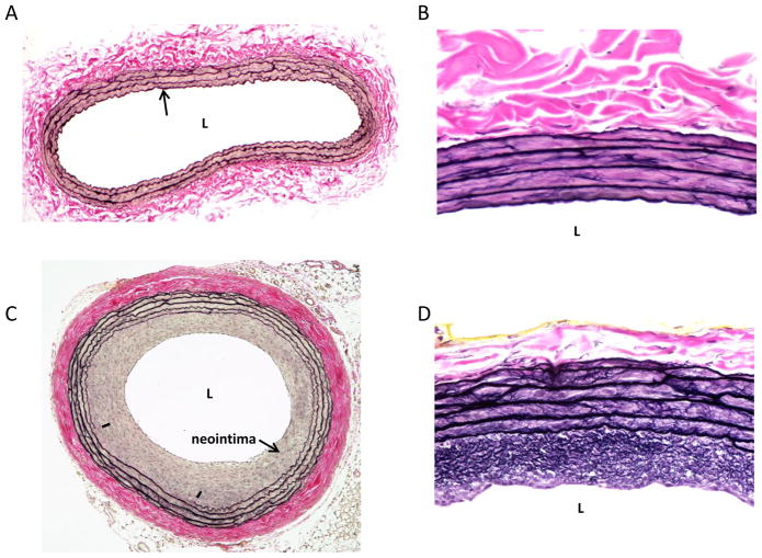
Representative Verhoeff-Van Gieson-stained cross-sections on rat left common carotid arteries. A: a whole cross-section of an uninjured artery is shown with a patent lumen (L), a single cell layer thick intima (arrow), a VSM-rich medial wall and a collagen-rich adventitia. B: a higher magnification photo of an uninjured artery with expanded details. C: shows a balloon-injured artery 2 weeks post-injury with a significantly reduced lumen and a robust concentric neointima. In this section a partially ruptured internal elastic lamina is noted (denoted by hash marks) along with a thickened and compacted collagen-rich adventitia. D: a higher magnification photo of an injured artery obtained 2 weeks post-injury clearly showing an elastin-rich neointima and enhanced medial wall elastin content. In all of these photomicrographs elastin fibers stain black (including the elastic laminae) and collagen and associated matrix components stain red (primarily the adventitia).

