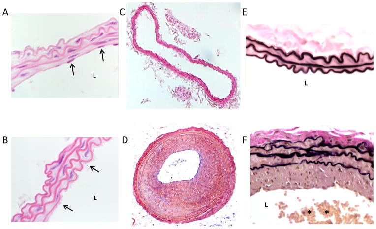
Figure 3.
Representative mouse carotid artery cross-sections with/without carotid artery wire denudation. A: a hematoxylin and eosin (H&E)-stained high magnification image of an uninjured carotid artery with nuclear staining of intimal endothelial cells (arrows) and a clear patent lumen (L). B: a H&E-stained high magnification image of a mouse carotid artery 30 minutes post-wire denudation injury. A platelet-rich monolayer covering the intimal lining is clearly evident (arrows). C: a H&E-stained cross-section of a mouse uninjured carotid artery and (D) a cross-section of a wire-injured carotid artery 4 weeks post-injury showing a robust and concentric neointima with severe luminal obstruction. E: a high magnification Verhoeff-Van Gieson-stained cross-section of a sham-operated (without wire denudation) mouse carotid artery 4 weeks post-sham surgery with a clear lumen and a thin intimal lining. F: a mouse Verhoeff-Van Gieson-stained carotid artery cross-section 4 weeks post-wire injury with significant neointima and a partially thrombosed, platelet-rich lumen (denoted by *).

