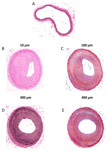
Figure 4.
Photomicrographs of mouse carotid artery cross-sections following complete blood flow obstruction via a common carotid artery ligation. A: an uninjured artery with clear lumen. B–E: carotid artery cross-sections obtained 4 weeks following common carotid ligation from the same mouse. Vessel shown in (B) was obtained 10 μm proximal to the site of ligation, (C) was obtained 100 μm proximal, (D) 300 μm and (E) 400 μm proximal to the site of ligation. It is noted that the degree of neointimal formation and the severity of stenosis is reduced the more proximal one moves away from the site of ligature.

