
FIG. 2.
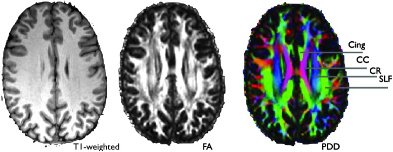
Example of diffusion tensor imaging-derived images that show the extra white-matter contrast gained by capturing diffusion anisotropy. The PDD (principal diffusion direction) map on the right is colored according to Red, left-right; Green, anterior-posterior; Blue, superior-inferior. Cing, cingulum bundle; CC, corpus callosum; CR, corona radiata, SLF, superior longitudinal fasciculus.