Fig. 1.
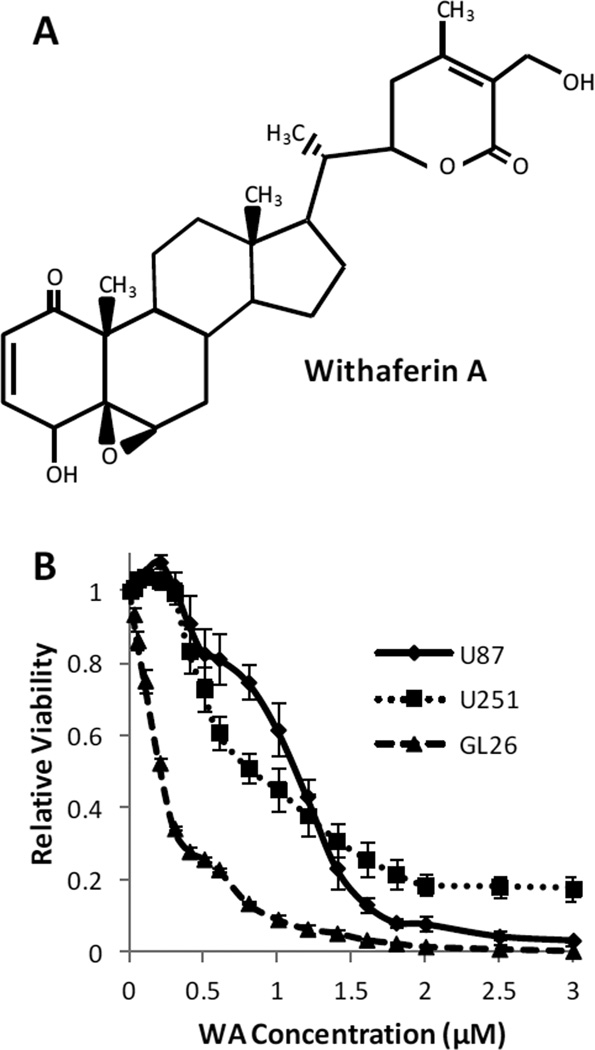
(A) Structure of withaferin A. (B) U87, U251, and GL26 cells were incubated with increasing concentrations of WA or a DMSO control for 72h and then assessed by MTS assay. WA dose escalation reduced cell proliferation and viability with IC50 values of 1.07±0.071µM, 0.69±0.041µM, and 0.23±0.015µM for U87, U251, and GL26 cells, respectively

