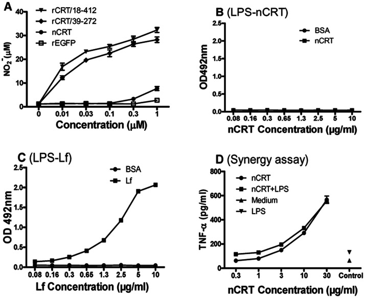
Figure 2. Lack of specific binding and sinergy between LPS and nCRT.
(A) Freshly isolated murine peritoneal macrophages were stimulated with rCRT/18-412, rCRT/39-272, nCRT or rEGFP (0.01–1 µM) for 24 hrs. Concentration of NO2 − in the culture supernatant was then determined using Griess Reagent and the results are expressed as mean NO2 − concentration (µM) ± SD. LPS-based ELISAs were performed for detection of LPS binding with CRT (B) or lactoferrin (C). Lf or nCRT (2 µg/ml) were added to wells in polyvinyl plates pre-coated with LPS (10 µg/ml), with BSA as a negative control. Combination of polyclonal rabbit Abs against CRT, or lactoferrin, and HRP-labeled goat-anti-rabbit IgG was used for detection with OPD as substrate. The results are expressed as absorbance at OD492 nm±SD. For sinergy analysis (D), freshly isolated mouse peritoneal macrophages were stimulated with nCRT (0.3–30 µg/ml) in the presence, or absence, of LPS (0.1 ng/ml) for 24 h. Cells in medium alone (Medium) or stimulated with LPS (0.1 ng/ml) alone (LPS) were included as controls. TNF-α in the culture supernatant was then quantitated using an ELISA kit and the results are expressed as mean concentration (pg/ml)±SD. These are representatives of 3 independent experiments.

