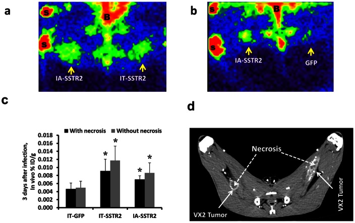
Figure 1. Imaging data of VX2 tumors in rabbits.
(a and b) Representative gamma camera planar images of VX2 tumors (a) infected in vivo by IA and IT administration of Ad-CMV-HA-SSTR2, and (b) infected in vivo by IA infusion of Ad-CMV-HA-SSTR2 and by IT injection of control Ad-CMV-GFP (B = bladder, S = source of 111In for positioning). Increased 111In-octreotide uptake is seen in tumors infected with Ad-CMV-HA-SSTR2 by both routes of administration compared to infection with the control Ad-CMV-GFP. (c) 111In-octreotide biodistribution in tumors normalized to tumor weight (%ID/g) calculated with and without necrosis using imaging only (in vivo biodistribution from gamma camera and CT imaging). Uptake was higher in tumors infected with Ad-CMV-HA-SSTR2 compared to control Ad-CMV-GFP (*, p<0.01, n = 6 for IA vs GFP; p<0.01, n = 6 for IT vs GFP). No difference was seen between the IA and IT groups. (d) Contrast enhanced CT showing the small amount of necrosis within the tumors.

