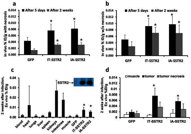
Figure 5. In vivo and ex vivo 111In-octreotide biodistribution.
(a and b) In vivo radioligand biodistribution normalized to tumor weight (%ID/g) calculated with (a) and without (b) necrosis at 5 days and 2 weeks after infection with Ad-CMV-HA-SSTR2 or control Ad-CMV-GFP (*, p<0.05 compared with GFP). Normalizing for necrosis demonstrates that the apparent loss of expression at the later 2 week time point (compared with 5 days) post infection is less when the confounding variable of necrosis is removed. (c) Graph showing ex vivo tissue biodistribution of 111In-octreotide in multiple organs in rabbits bearing VX2 tumors 2 weeks post virus infection. Significantly greater uptake was seen in tumors infected with Ad-CMV-HA-SSTR2 by IA or IT routes compared to tumors infected with control Ad-CMV-GFP (c and d: *, p<0.001 for IA, n = 6; p<0.01 for IT, n = 4) and this is confirmed by Western blotting. (d) Graph showing the %ID/g was significantly higher in viable tumor tissue infected with Ad-CMV-HA-SSTR2 compared to tumor necrosis (p<0.01 for IA, n = 6, p<0.02 for IT, n = 4) or muscle (p<0.001 for IA, n = 6, p<0.02 for IT, n = 4) in tumors infected by Ad-CMV-HA-SSTR2, but not control Ad-CMV-GFP.

