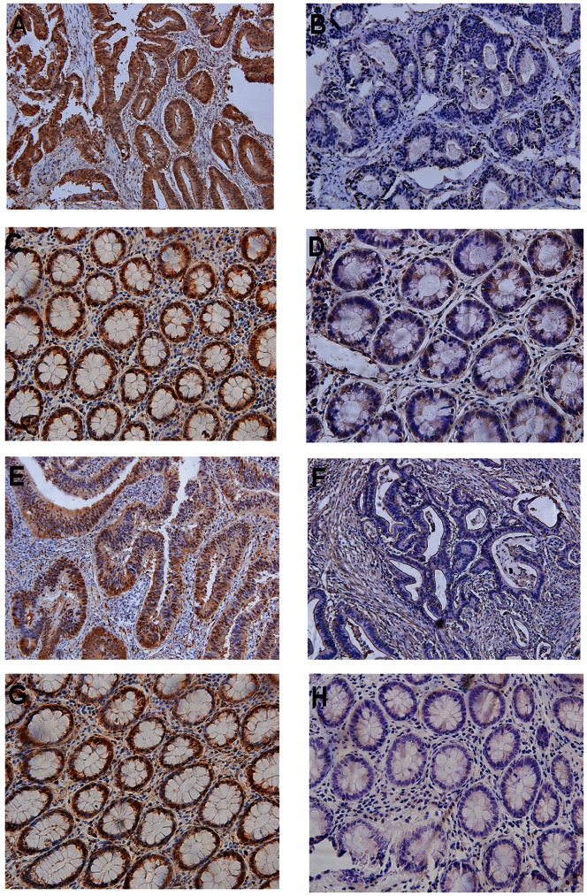
Figure 1. Immunohistochemically stained tissues from CRC patients and adjacent normal control tissues.
(A) YAP-positive tumor; (B) YAP-negative tumor; (C) YAP-positive normal tissue; (D) YAP-negative normal tissue; (E) TAZ-positive tumor; (F) TAZ-negative tumor; (G) TAZ-positive normal tissue; (H) TAZ-negative normal tissue. Representative images were taken under a microscope (x10). These results indicate the clinical significance of YAP and TAZ overexpression in CRC tissue.

