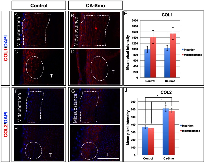
Figure 2. Expression of collagens in control and constitutively active Smo (CA-Smo) patellar tendons at E17.5.
Panels A–D show immunohistochemistry of COL1 in control (A and C) and CA-Smo (B and D) sagittal sections of knee joints. Panels F–I show immunohistochemistry of COL2 in control (F and H) and CA-Smo (G and I). Activation of Smo in the tendon does not affect the expression of COL1 but causes ectopic expression of COL2 in the midsubstance. Panel E and J show pixel intensity measurements of COL1 and COL2 staining. COL2 was up-regulated in the CA-Smo mice throughout whole tendon. White dotted line marks the region of midsubstance. White dotted circle marks the insertion site. Error bar represents standard error and the asterisk represents statistical significance (p<0.05). Blue staining shows nuclear staining using DAPI. T = Tibia. Scale bar = 100 µm.

