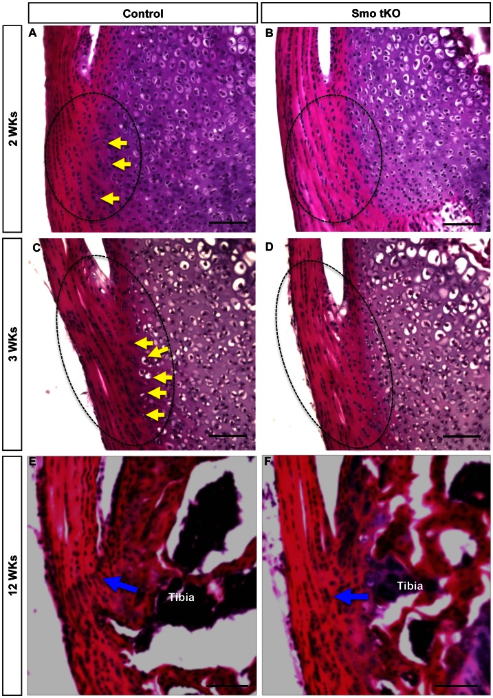
Figure 6. H&E staining of the tibial insertion site of patellar tendon of control and ScxCre;Smof/−(Smo tKO).
Panel A–B show H&E staining of the sagittal sections of 2 weeks (2 wks) old control (A) and Smo tKO (B). Panel C–D show H&E staining of the sagittal sections of 3 wks old control (C) and Smo tKO (D). Panel E–F show H&E staining of the sagittal sections of 12 wks old control (E) and SmotKO (F). There are fewer chondrogenic cells (yellow arrow) in the tibial insertion site of patellar tendons in Smo tKO animals, and the tidemark of the Smo tKO mice is absent. See text for details. Yellow arrow = advanced differentiated cells. Blue arrow = tidemark. Scale bar = 100 µm.

