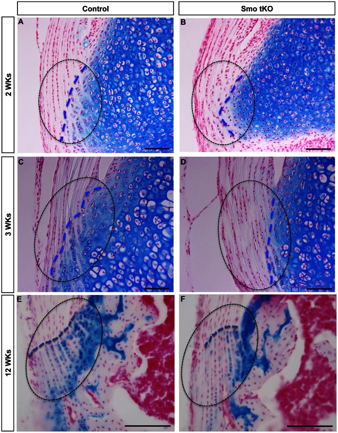
Figure 7. Alcian blue staining is decreased in the tibial insertion sites of patellar tendons of ScxCre;Smof/−(Smo tKO).
Shows alcian blue staining in insertion sites of control (A, C, and E) and Smo tKO (B, D, and F) patellar tendons at 2 wks (A and B), 3 wks (C and D) and 12 wks (E and F). See text for details. Dotted line outlines the insertion site. Blue dotted line shows the boundary between the tendon and tibial cartilage. See text for details. Cell nuclei are stained using fast red. Scale bar = 100 µm.

