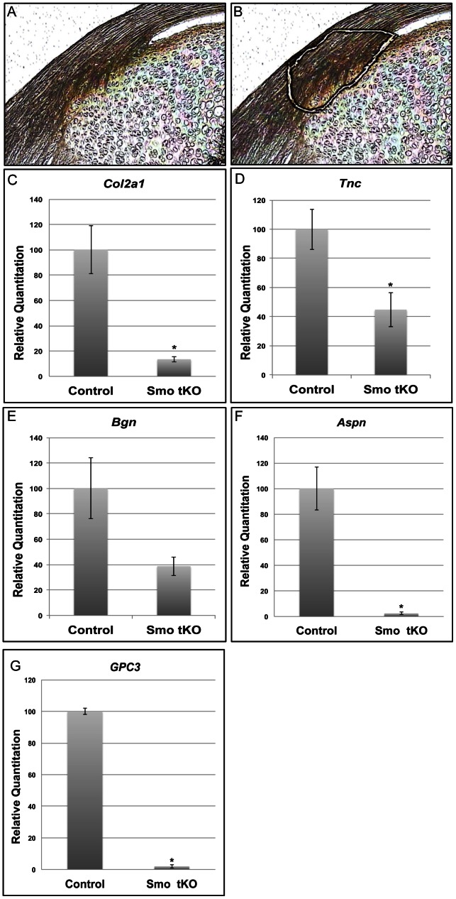
Figure 8. Expression of insertion site markers in the tibial insertion site of patellar tendon of ScxCre;Smof/−(Smo tKO).
Panels A–B show a mid-sagittal section of 2 wks old mouse patellar tendon before (A) and after (B) LCM dissection of the insertion site. Panels C–G show Q-PCR results of Col2a1(C), Tnc (D), Bgn (E) Aspn (F) and Gpc3 (G). All these markers were reduced in the Smo tKO animals. See text for details. The asterisk represents statistical significance (p<0.05).

