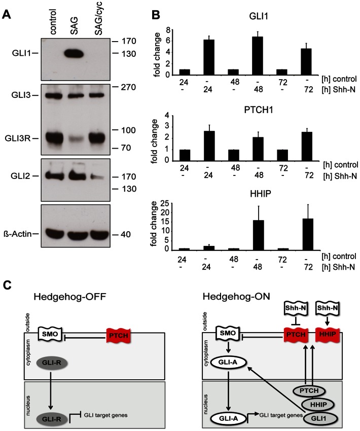
Figure 1. Daoy cells respond to SAG and Shh-N by upregulating canonical HH/GLI targets.
(A) Incubation with SAG (100 nM) induced the expression of GLI1 protein and inhibited processing of endogenous GLI3 to its repressor form. Co-incubation with Cyclopamine (cyc, 5 µM) inhibited the SAG-induced GLI1 expression and promoted processing of GLI3 to its repressor (GLI3R) form and inhibited GLI2 expression. Beta-actin shown as Western blot loading control. (B) Transcripts for GLI1 and PTCH were upregulated after stimulation with Shh-N for 24 h, induction of HHIP transcripts was seen after 48 h. Enhanced GLI1, HHIP, and PTCH expression levels were still observed after 72 hours. (C) Schematic presentation of canonical Hedgehog signaling. Left panel illustrates silencing of Hedgehog-mediated signaling via a PTCH-mediated block of SMO so that repressor GLI3 prevails and limits the expression rate of Hedgehog target genes. The right panel illustrates the “Hedgehog-on” state, activating GLI proteins now control the expression of Hedgehog-target genes such as GLI1, PTCH, and HHIP. Upregulation of PTCH and HHIP will eventually result in the downregulation of Hedgehog-signaling. Red color indicates a protein with inactivating properties, white color indicates a protein with activating properties, and grey color indicates RNA transcripts.

