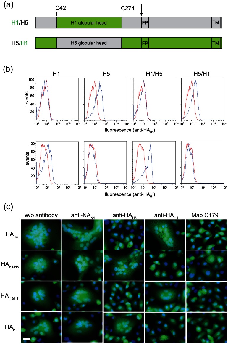
Figure 4. Inhibition of HA-mediated syncytia formation by chicken immune sera.
(a) Diagrams of chimeric hemagglutinins. The chimeric H1/H5 hemagglutinin was constructed by swapping the globular head domain located between C42 and C274 of A/chicken/Yamaguchi/8/2004 (H5N1) HAH5 (grey) with that of the corresponding region from A/duck/Italy/1447/2005 (H1N1) HAH1 (green). The chimeric H5/H1 hemagglutinin was constructed accordingly by replacing the HAH1 globular head domain with the corresponding HAH5 domain. The proteolytic cleavage site (arrow), fusion peptide (FP) and transmembrane (TM) domain are indicated. (b) Flow cytometric analysis of BHK-21 cells expressing parental and chimeric HAs. Cells were infected with either VSV*ΔG (blue graphs) or VRPs expressing the indicated HAs (red graphs). At 6 hours p.i., cells were stained with chicken immune sera specific for either HAH5 or HAH1 and goat anti-chicken IgY Alexa-546 conjugates. (c) Inhibition of syncytia formation. Vero cells were infected with VRPs expressing the indicated HAs using an MOI of 5 ffu/cell. At 5 hours p.i., cells were treated for 60 minutes with acetylated trypsin to proteolytically activate HAH1 and HAH5/H1. At 6 hours p.i., the cells were incubated for 30 minutes at 37°C with the indicated chicken immune sera (diluted 1∶20 in MEM), exposed for 5 minutes at 37°C to pH 5.4, further incubated in medium for 2 hours, and fixed with paraformaldehyde. The nuclei were stained with DAPI (blue fluorescence). Expression of eGFP is indicated by green fluorescence. Scale bar represents 20 µm.

