Fig. 3.
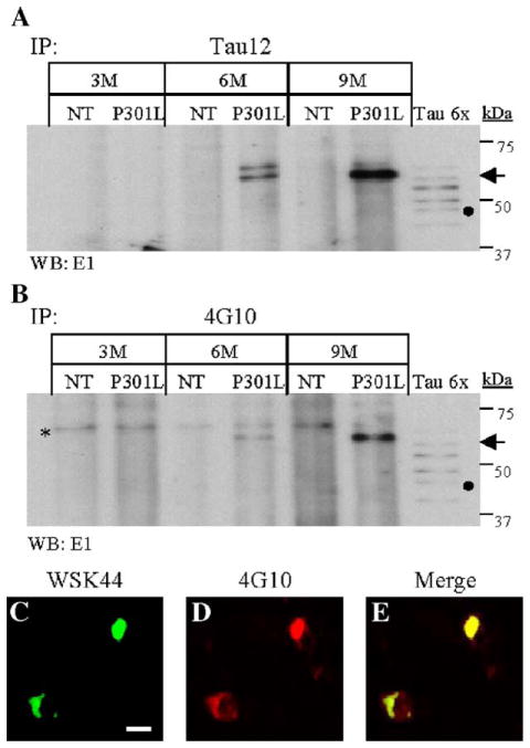
Sarkosyl-insoluble tau and tau inclusions display phosphotyrosine immunoreactivity. Tau proteins present in the sarkosyl-insoluble fraction of NT and JNPL3 (P301L) mouse brains, from 3, 6, and 9 months of age, were immunoprecipitated (IP) using either Tau12 (A) or 4G10 antibody (B). (A –B) IP tau proteins were detected by Western blot analysis using E1 antibody. The lanes designated Tau6x contain six isoforms of recombinant tau. The filled circle on panels A and B marked the potion of the 0N4R tau isoform. The arrows indicate the 64-kDa tau species in panels A and B. The asterisk on panel B marks cross-reacting labeled band in both NT and P301L samples. (C –E) Spinal cord transverse sections from a 10-month-old JNPL3 (C– E) mice were used for immunofluorescence analysis using tau-specific antibody WSK44 (C-green) and phospho-tyrosine-specific antibody 4G10 (B-red). Neurons immunoreactive to both antibodies display yellow signal (E-merge). The cells were visualized by confocal microscopy at 0.1 μm. The scale bar in panel C = 100 μm.

