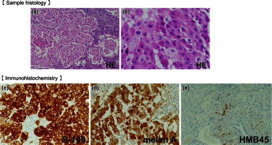
Fig. 8.

H&E staining revealed stranded proliferation of spindle-shaped cells and higher magnification views showed bright cytoplasm and nuclear atypia (a lower magnification; b higher magnification). Immunohistochemistry: c negativity for HMB-45; d positivity for S-100 protein; and e positivity for Melan A. Deposition of brownish black pigmented granules, usually recognized in melanotic melanoma, was not observed
