
Figure 3. Electrophysiology of direction-specific uniform DC EFs in synaptic pathways of the rat motor cortex.
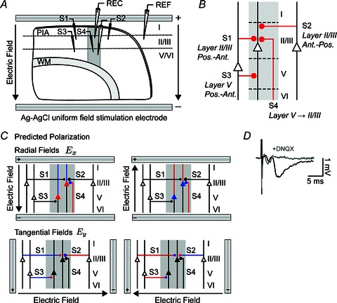
A, schematic of electrophysiology setup where uniform extracellular EFs were generated in all experiments by passing constant current across parallel Ag–AgCl wires positioned in the bath across the slice. Activity was monitored in layer II/III or layer V with a glass microelectrode. An additional field electrode (REF) was positioned in an iso-potential to remove the uniform field artifact. Activity was evoked with a bipolar stimulating electrode (S1–S4) positioned 500 μm from the recording electrode in either layer II/III or layer V targeting one of four distinct synaptic pathways corresponding to different orientations of afferent axons: posterior horizontal layer II/III (S1); anterior horizontal layer II/III (S2); posterior horizontal layer V (S3); and vertical layer V to II/III (S4). B, diagram summarizing the primary synaptic circuits in this study. Line thicknesses and diameters of the filled circles, which represent synapses, are correlated with the strength of the synaptic input. C, schematic of the expected polarization in distinct synaptic pathways exposed to radial and tangential fields. Somas, dendrites, axons and axon terminals are depolarized (red), hyperpolarized (blue) or not affected (black) by DC fields. D, characteristic fEPSP and field spike waveforms from the layer V pathway. The fEPSPs, but not earlier field spike, were suppressed by the non-NMDA receptor antagonist 6,7-dinitroquinoxaline-2,3-dione (DNQX). WM, white matter.