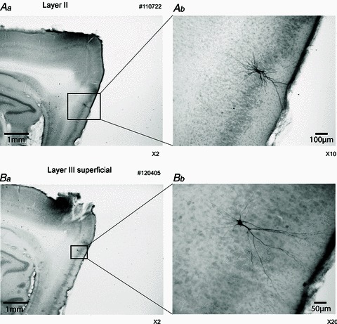
Figure 2. Examples of the recorded cells.

Aa, layer II cell (parasagittal section). Ab, higher magnification of Aa. The biotin-labelled cell is a stellate cell in layer II of the medial entorhinal cortex (MEC). Ba, layer III superficial cell (parasagittal section). Bb, higher magnification of Ba. This neuron shows non-spiny dendrites, and the initial bifurcation is close to the cell body, which indicates a type 2 projection neuron as described by Gloveli et al. (1997).
