Fig. 1.

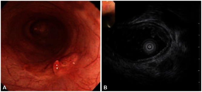
Esophageal carcinoma with submucosal invasion. (A) Endoscopy shows flat nodular elevated mass. (B) Hypoechoic mass in the mucosal layer with the third layer thinning is seen on endoscopic ultrasonography.

Esophageal carcinoma with submucosal invasion. (A) Endoscopy shows flat nodular elevated mass. (B) Hypoechoic mass in the mucosal layer with the third layer thinning is seen on endoscopic ultrasonography.