
Fig. 3.
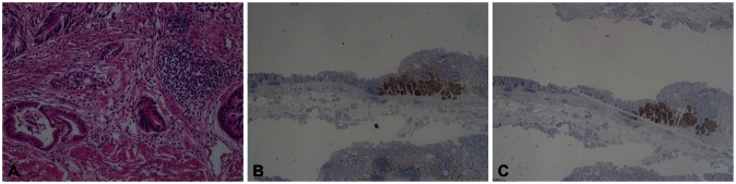
Pathologic findings. (A) It shows a well-differentiated adenocarcinoma and a neuroendocrine carcinoma. One area of transition between the two histologic types was observed (H&E stain, ×200). (B) The tumor cells with endocrine differentiation are highlighted by CD 56 stain, but adenocarcinoma shows no immunoreactivity (CD 56 stain, ×40). (C) The tumor cells with endocrine differentiation are highlighted by synaptophysin stain, but adenocarcinoma shows no immunoreactivity (synaptophysin stain, ×40).