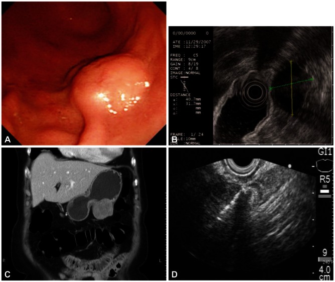
Fig. 1.
(A) Gastroscopy shows a submucosal elevated lesion with bridging fold. (B) Endoscopic ultrasound (EUS) shows a 4 cm-sized hypoehoic mass with internal hyperechogenicity originating from the proper muscle layer. (C) Contrast enhanced computed tomography scan shows a round, enhancing extraluminal-growing mass. (D) EUS-guided Trucut biopsy with 19 gauze needle was performed.

