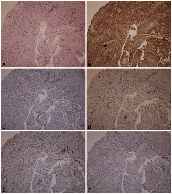
Fig. 2.
Pathologic findings of Trucut biopsy. Tissue was composed of broad bundles of elongated cells (A, H&E stain, ×100). Immunohistochemistry (IHCS) findings; (B) the tumor strongly stains for S-100 (arrow). IHCS staining for (C) CD 117, (D) CD 34, (E) smooth muscle actin, and (F) desmin are negative (H&E stain, ×100).

