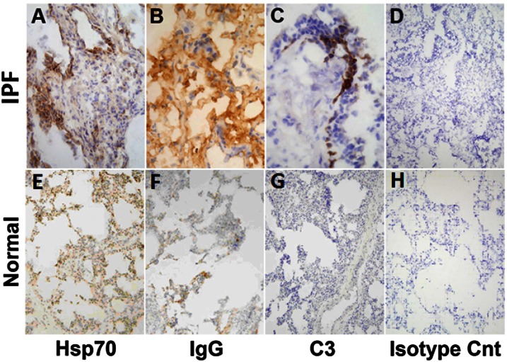
Figure 2.
Lung immunohistochemistry. Columns from left to right depict, respectively, expression of heat shock protein 70 (HSP70), IgG immune complexes (IgG), complement deposits (C3), and isotype controls. Rows, from top to bottom respectively, depict end-stage idiopathic pulmonary fibrosis (IPF) lungs explanted during therapeutic pulmonary transplantations and normal lungs harvested during multiorgan retrievals but not used in therapeutic transplantations (n = 6 each). IPF specimens were depicted at 20× to better illustrate anatomical localizations of the HSP70, IgG, and C3. Normal lung sections are shown at 10× to optimally depict the overall paucity of the expressions/depositions in these specimens.

