Fig. 2.
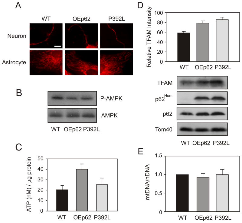
p62 overexpression increases the functionality of mitochondria in the hippocampus. A) Mitochondrial morphology was altered in OEp62 mice compared to WT and p62:P392L. Primary neuronal cells were cultured from dissected embryonic Day 19 hippocampus. At culture day 7, mitochondria were visualized by staining with MitoTracker Red and examined using a confocal microscope. B) Activated AMPK levels were decreased in p62 overexpressing tissue. Hippocampal lysates from WT, OEp62 and p62:P392L mice were separated on SDS-PAGE and phospho-AMPK levels compared to total AMPK examined by Western blot. C) Overexpression of p62 results in increased ATP production. Total ATP levels in hippocampal lysates were measured by luciferase assay and compared to WT. D) Mitochondrial import was increased with p62 overexpression. Mitochondria were isolated from hippocampal lysates and import of TFAM examined by SDS-PAGE and Western blot. Presence of overexpressed protein was confirmed with p62Hum antibody and compared to native protein. E) p62 overexpression did not show increased mtDNA levels. Total mitochondrial DNA copy number was quantitated by RT-PCR.

