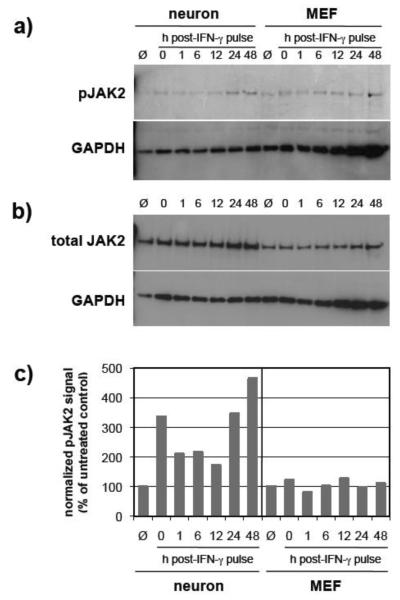
Figure 3. JAK2 phosphorylation in IFN-γ-treated neurons is significantly extended as compared to MEF.
Neurons and MEF were treated as described (Figure 1), and equal volumes of cell lysates were examined by immunoblotting for a) phospho-JAK2 (pY1007/1008); b) total JAK2; and GAPDH signals. c) Blot signals were quantified using densitometry, and phospho-JAK2 signals were normalized to total JAK2 expression and to GAPDH for loading. Values are expressed as a percent of control. Data shown are from a representative experiment. Ø: cells not exposed to IFN-γ.

