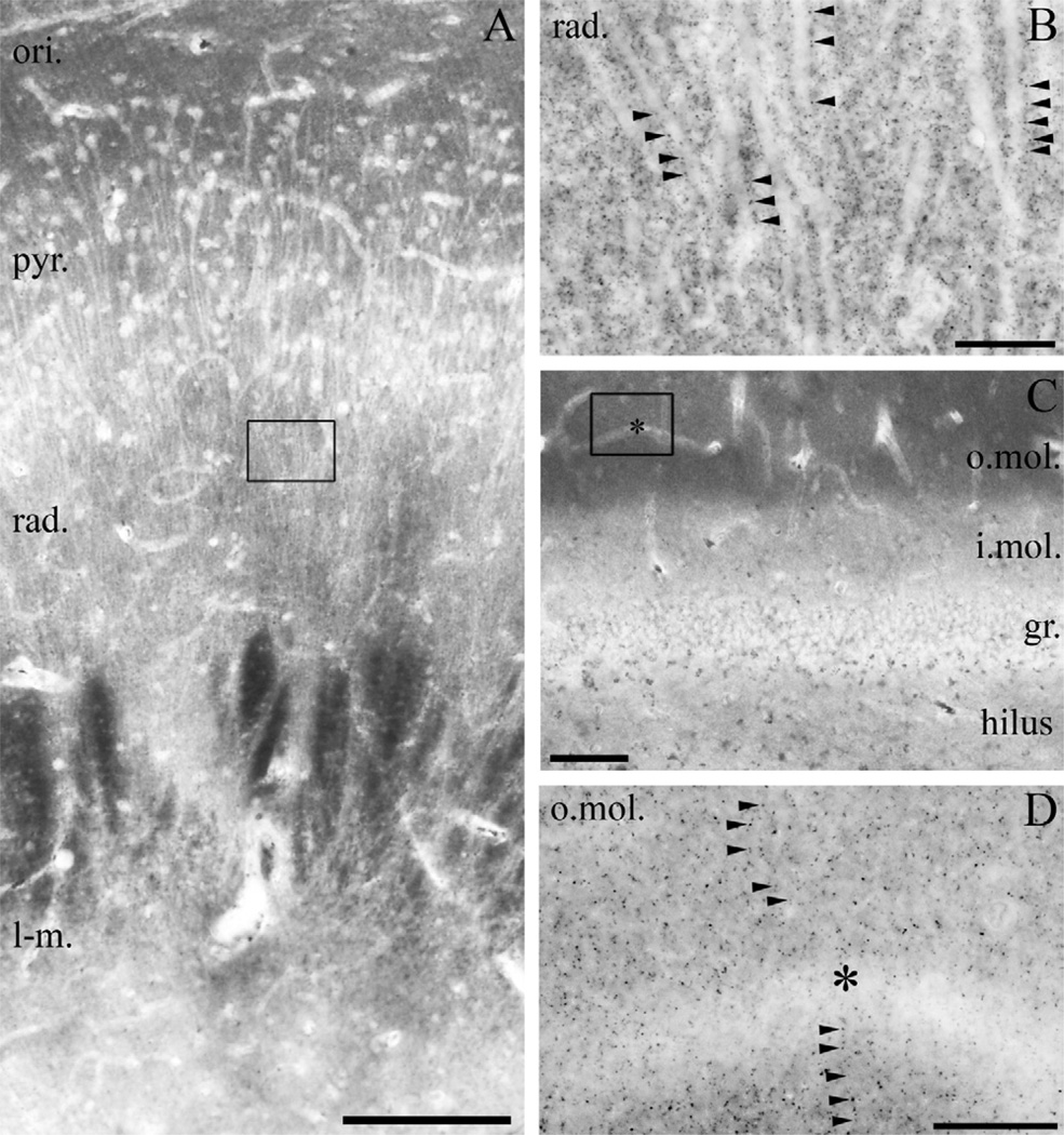
Fig. 5.
Light microscopic localization of MGL in the human hippocampus. (A) Light micrograph of immunoperoxidase staining for MGL in the CA1 subfield of the cornu ammonis shows that the highest density of MGL-immunoreactivity using the “MGL-mid” antibody is located in the stratum oriens (ori.), but significant levels of immunostaining is also apparent in strata pyramidale (pyr.), radiatum (rad.) and lacunosum-moleculare (l-m.). (B) At higher magnification, the abundant neuropil staining becomes visible, e.g. in the stratum radiatum of CA1 region (depicted here). The characteristic MGL-immunopositive granules (arrowheads) decorate MGL-immunonegative apical dendrites of pyramidal cells. (C) In the dentate gyrus, MGL-immunostaining was observed predominantly in the outer two thirds of the stratum moleculare (o.mol.), whereas it was weaker in the inner third of stratum moleculare (i. mol.), stratum granulosum (gr.) and in the hilus. Somata of granule cells (similarly to somata of pyramidal cells in A) are hardly or not at all labeled by MGL immunostaining. (D) At higher magnification, the punctate neuropil staining is present throughout the stratum moleculare of the dentate gyrus. MGL-immunonegative dendrites are frequently outlined by the characteristic MGL-containing varicosities often arranged in an array-like manner. Open boxes denote the positions of corresponding insets (B in A, D in C). Asterisks in (C–D) depicts the same capillary used as landmark. Scale bars: (A) 200 µm; (B, D) 20 µm; (C) 100 µm.

