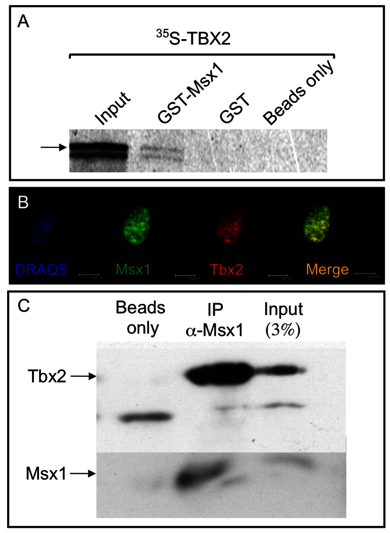
Fig. 2.

Msx1 and Tbx2 proteins can physically interact in vitro. (A) GST-Msx1 was able to pull down 35S-labeled Tbx2 (arrow; lane 2 from left). As a control, the GST moiety (lane 3) or beads alone (lane 4) were not able to pull down any Tbx2. (B) Msx1 and Tbx2 are endogenously expressed in C3H10T1/2 cells. The nucleus was stained using DRAQ5. (C) Co-immunoprecipitation was carried out in C3H10T1/2 cells as indicated. Tbx2 and Msx1 were assayed following immunoprecipitation with anti-Msx1 antibody. Whereas protein A beads alone did not show any Tbx2 (lane 1 from left), in the presence of anti-Msx1 antibody there is robust presence of Tbx2 (lane 2). As a control, Tbx2 and Msx1 are detected in the input sample (lane 3).
