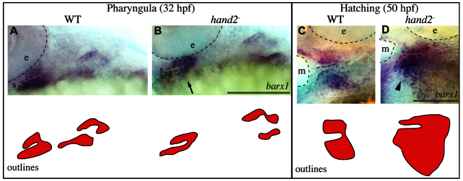
Fig. 8.

hand2 regulates barx1 positively and negatively early and late, respectively. (A-D) barx1 expression was revealed by in situ hybridization and imaged with transmitted light. Labeled pharyngula period anatomy includes the stomodeum (s) and the eye (e). Labeled hatching period anatomy includes the mouth (m) and the eye (e). Arrow and arrowhead indicate reduced and expanded ventral barx1 expression, respectively. Scale bars: 100 μm.
