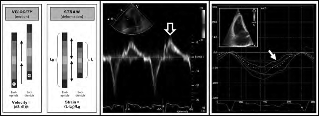
Figure 1. Advanced echocardiographic techniques for the evaluation of cardiac involvement in systemic sclerosis: tissue Doppler imaging vs. speckle-tracking echocardiography.
Left panel: Differences between myocardial velocity and myocardial strain. Middle panel: Example of tissue velocities of basal RV free wall in a patient with SSc [open arrow points to peak systolic (S’) tissue velocity]. Right panel: Example of systolic strain curves of the RV derived from speckle-tracking echocardiography in the same patient with SSc (inset = 2D speckle-tracking image; arrow points to peak systolic strain of the basal RV free wall). In this case, peak systolic strain (−4.4%) was reduced (consistent with intrinsic RV systolic dysfunction) whereas peak systolic tissue velocity (12.3 cm/s) was preserved. RV systolic tissue velocity was normal despite abnormal RV function due to the tethering effect of normal LV systolic function (i.e. RV longitudinal velocity, but not strain, is somewhat dependent on LV systolic function as the LV ‘pulls’ the RV base toward the apex during systole). Thus, speckle-tracking strain analysis may be advantageous (compared with tissue Doppler imaging) for the detection of subclinical cardiac involvement.

