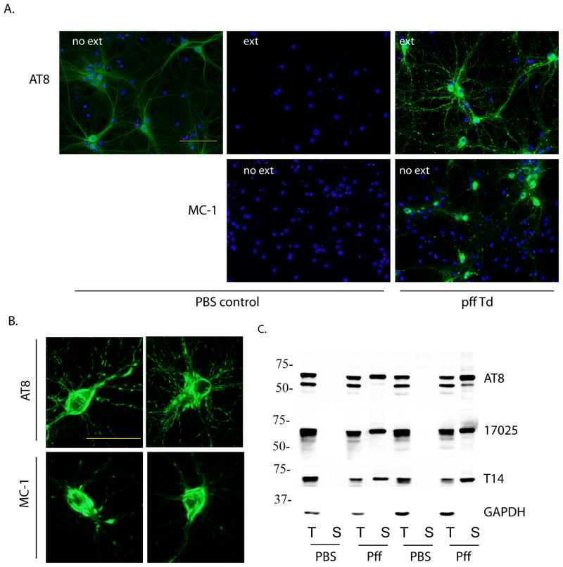
Figure 1. Insoluble tau accumulated in PS19 primary hippocampal neurons after incubation with myc-K18/P301L pffs.
(A) PS19 neurons treated with PBS (PBS control) or myc-K18/P301L fibrils (pff Td) were immunostained with phospho-tau mAb AT8 and conformational mAb MC-1 with or without 1% Triton-X100 extraction during fixing (ext, no ext, respectively). DAPI staining was used to visualize nuclei (blue). Scale bar: 100 μm. (B) Perikaryal tau aggregates recognized by AT8 and MC-1. Scale bar: 50 μm. (C) Neurons treated with PBS or myc-K18/P301L fibrils (pff) were sequentially extracted with 1% Triton-X100 lysis buffer (T) followed by 1% SDS lysis buffer (S) and immunoblotted with AT8, polyclonal antibody against total tau (17025), and mAb against human tau (T14). GAPDH served as loading control. Results from two independent sets of neurons were shown.

