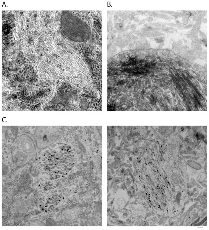
Figure 2. Ultrastructural analysis of tau pff-induced aggregates by EM and immune-EM.
Routine EM (A) revealed filamentous structures in the cytoplasm of tau pff transduced neurons, which were recognized by MC-1 in immuno-EM using both HRP-labeling (B) and nanogold-amplification (C) detecting methods. Scale bar: 500 nm.

