Fig. 2.
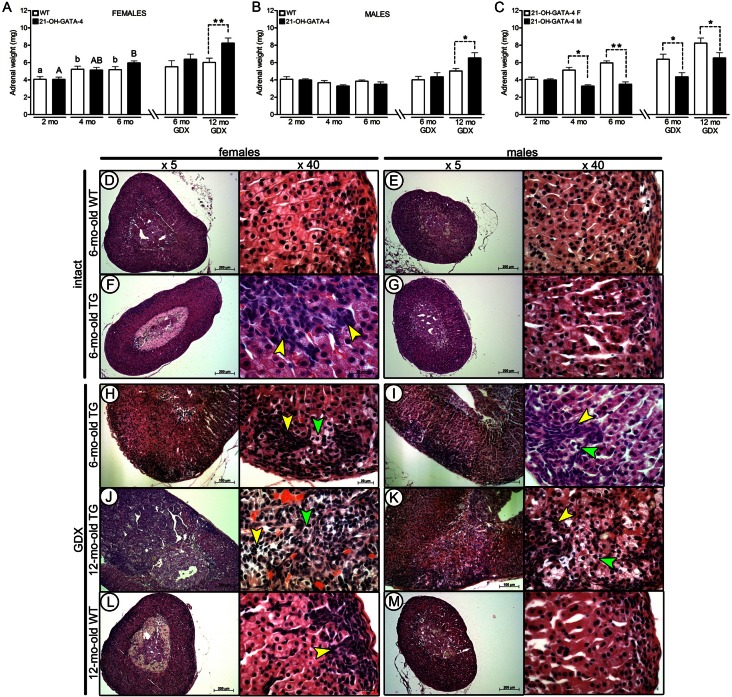
Weights and morphological characteristics of intact and GDX WT and 21-OH-GATA-4 adrenals. (A–C) Total adrenal weights (mean±s.e.m.) of WT and TG female and male mice (n = 16, 12 and 8 for the TG, WT and GDX groups, respectively). Different letters above the bars indicate significant differences between groups (small letters and white bars for females, capitals and black bars for males). Asterisks indicate differences between designated groups (*P<0.05; **P<0.01). ND, non-detectable; TG; transgenic 21-OH-GATA-4 mice; GDX, gonadectomy/gonadectomized; WT, wild type. (D–M) Histopathology of intact 6-month-old TG female (F), male (G) and control littermate (D,E) adrenals. In adrenal cortex of 6-month-old TG females, intensively stained spindle-shaped (A-type, yellow arrowheads) neoplastic cells originating from the subcapsular region are seen (F). Both 6-month-old GDX TG females (H) and males (I) developed multiple hyperplastic foci of invasive A cells by the age of 6 months (small with spindle-shaped nucleus; yellow arrowheads) and large single B cells (larger, with pale cytoplasm; green arrowheads). Post-GDX adenomas were induced in 12-month-old TG adrenals (J,K) that were not observed in control littermates (L,M).

