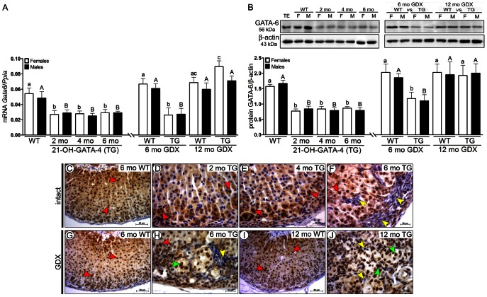
Fig. 3.
GATA-4 regulated spatiotemporal expression of GATA-6 in adrenals of 21-OH-GATA-4 mice. (A) Quantification of Gata6 mRNA by qPCR. Each bar represents the mean±s.e.m. (n = 5) relative to Ppia. (B) Western blot of GATA-6 protein in 2-, 4- and 6-month-old intact and 6- and 12-month-old GDX TG and WT mice. The upper panels show representative western blots of GATA-6 and β-actin. Murine WT testis (TE) was used as a positive control. The lower panels show densitometric quantification of the bands. Each bar represents the mean±s.e.m. (n = 5) relative to β-actin. Different letters above the bars indicate significant differences between groups (small letters and white bars for females, capitals and black bars for males). ND, non-detectable; TG, transgenic 21-OH-GATA-4 mice; F, females; M, males; GDX, gonadectomy/gonadectomized; WT, wild type; TE, testis. (C–J) Immunohistochemistry of GATA-6 in adrenal glands of 2-, 4- and 6-month-old intact TG and WT females (C–F), and in 6- or 12-month-old GDX TG and WT females (G–J). Clear lack of nuclear immunoreactivity for GATA-6 was observed in neoplastic A cells (yellow arrowheads) of intact (F) and GDX (H,J) TG adrenals in comparison with normal adrenocortical cells (C,D; red arrowheads). In 6- and 12-month-old GDX TG female adrenals, expression of GATA-6 was observed, throughout the histologically normal adrenal cortex and in B cells (F,J; green arrowheads).

