Fig. 6.
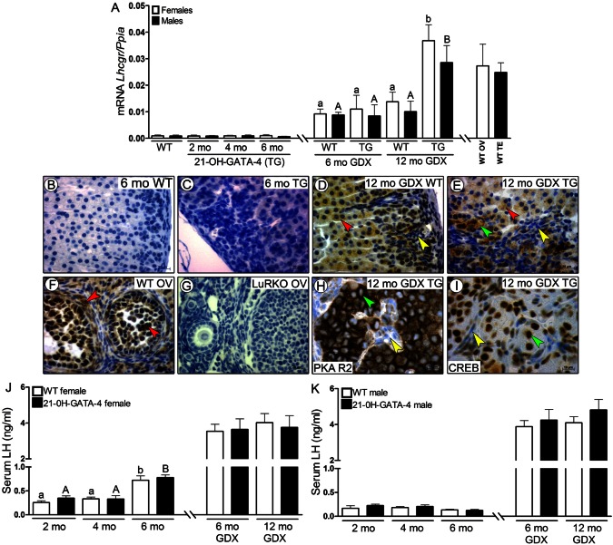
Expression of LHCGR and activation of PKA R2 and CREB in 21-OH-GATA-4 adrenals, as well as serum LH concentration in 21-OH-GATA-4 mice. (A) qPCR analysis of Lhcgr mRNA expression in adrenal glands of intact (2-, 4- and 6-month-old) and GDX (6- and 12-month-old) TG and WT mice. Each bar represents the mean±s.e.m. relative to Ppia expression (n = 5 per group). Different letters above the bars indicate significant differences between groups (small letters and white bars for females, capitals and black bars for males). TG, transgenic 21-OH-GATA-4 mice; GDX, gonadectomized, WT, wild type; OV, ovary; TE, testis. (B–I) Immunohistochemistry of LHCGR in adrenal glands of 6-month-old intact and 12-month-old GDX TG females (C,E) and their control littermates (B,D). WT and LuRKO (luteinizing hormone receptor knockout mice) ovaries were used as positive and negative controls, respectively, for the LHCGR antibody (F,G). Adrenal glands of intact TG and WT mice were LHCGR negative (B,C). Positive staining for LHCGR was observed in normal cortex (red arrowheads) and in B cells (green arrowheads) but not in A cells (yellow arrowheads) of GDX WT and TG mice (D,E). LHCGR in WT ovary was localized to granulosa, theca and luteal cells (F, red arrowheads), whereas all structures of the LuRKO ovary were LHCGR negative (G). LHCGR-positive B cells, but not A cells, were PKA R2 (H) and CREB positive (I) in 12-month-old GDX TG adrenals. (J,K) Serum LH concentration in 2-, 4- and 6-month-old intact, and 6- and 12-month-old GDX TG and WT mice. Each bar represents the mean LH concentration (±s.e.m.) of at least eight individuals in each group.

