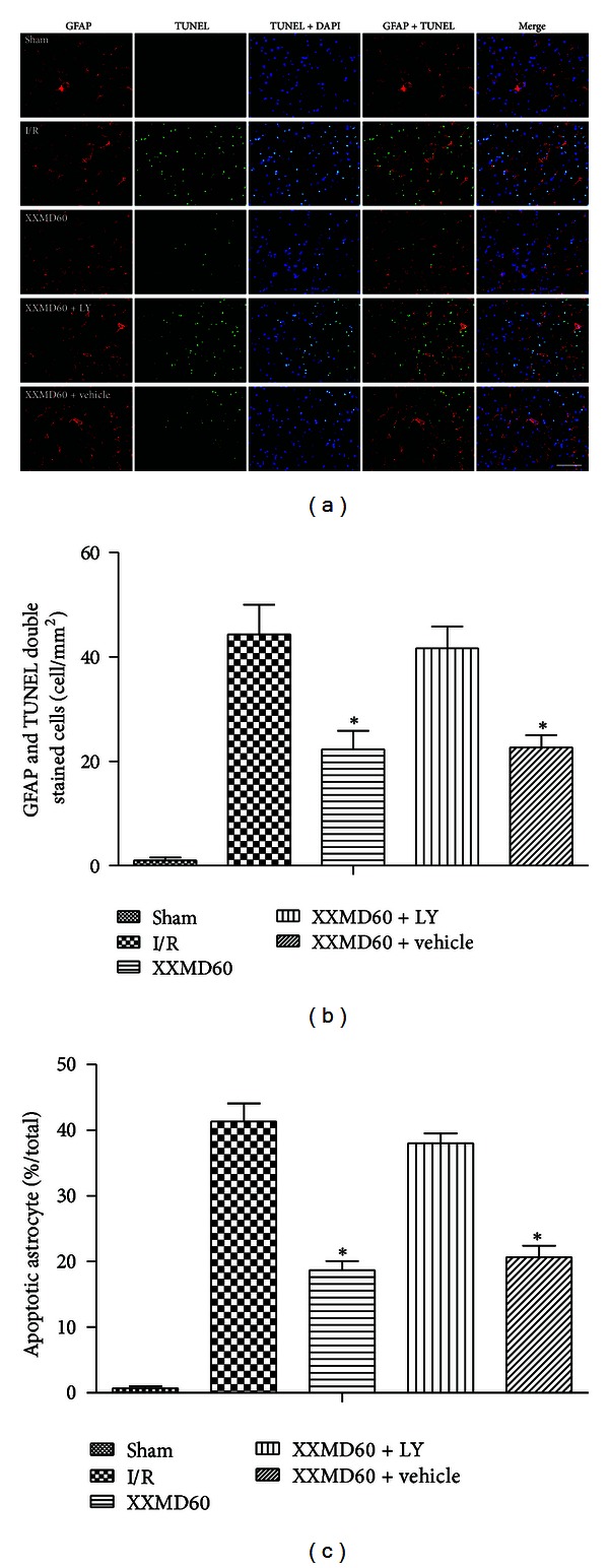
Figure 5.

Representative images of astrocyte apoptosis in penumbra of ischemic cortex at 24 h after reperfusion. (a) Representative photomicrographs of immunofluorescence labeling with GFAP (red) and TUNEL (green) double staining. Nuclei were counterstained with DAPI (blue). And collocation of green and blue indicated TUNEL positive cell in the same view. Stroke with reperfusion notably caused TUNEL-positive cells at 24 h after reperfusion without XXMD treatment, and a decrease was found in XXMD60 group. However, LY294002 abolished the reduction. (b) Quantification of apoptotic astrocytes. TUNEL and GFAP double stained cells (yellow) indicated the apoptotic astrocytes. A marked increase in apoptotic astrocytes was found at 24 h after reperfusion and was reversed by XXMD treatment. (c) Percentage of apoptotic astrocyte in GFAP positive cell. There were more nonapoptotic astrocytes in the XXMD-treated groups in absence of LY294002 compared with the I/R group. LY, LY294002. Scale bar = 50 μm. Data are reported as the means ± SEM. n = 6; *P < 0.05 versus the I/R group.
