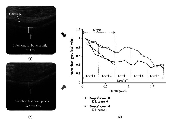
Figure 1.

(a) Ultrasound image of healthy knee cartilage-bone interface. A rectangular bone segment was selected in location perpendicular to the incident ultrasound beam. (b) Ultrasound image of osteoarthritic knee cartilage-bone interface. (c) Comparison of nonosteoarthritic (black) and osteoarthritic (gray) subchondral bone gray-level intensity profiles demonstrating decreasing subchondral bone reflection with depth. Five uniform depth levels, overall bone level and slopes calculated for first 2 levels are marked.
