Abstract
Reverse genetics approaches that enable the generation of recombinant influenza A viruses entirely from plasmids are invaluable for studies on virus replication, morphogenesis, pathogenesis, or transmission. Furthermore, influenza virus reverse genetics is now critical for the development of new vaccines for this human and animal pathogen. Periodically, influenza gene segments are unstable within plasmids in bacteria. The PB2 gene segment of a highly pathogenic avian H5 influenza virus A/Turkey/Ontario/7732/1966 (Ty/Ont) was unstable in commonly available cloning plasmids (e.g., pcDNA3.1/V5-His-TOPO) and in standard influenza virus reverse genetics plasmids (e.g., pHH21), which contain high copy origins of replication. Thus, a low-copy influenza reverse genetics plasmid (pGJ3C3) was developed to enable rapid cloning of unstable influenza A virus genes using ligation-independent recombination-based cloning. The unstable Ty/Ont PB2 gene segment was efficiently cloned using the pGJ3C3 plasmid and this clone was used to rescue a recombinant Ty/Ont virus. This low copy reverse genetics plasmid will be useful for cloning other unstable segments of influenza A viruses in order to rescue recombinant viruses, which will facilitate basic studies and vaccine seed stock production.
Keywords: Influenza, reverse genetics, ligation-independent cloning, recombination-based cloning, low copy plasmid, unstable gene
Influenza A virus is an important pathogen that infects many different species, including human, swine, equine, canine, and avian (Wright et al., 2007). Annual influenza epidemics infect 250–500 million people, resulting in 250–500 thousand fatalities. Occasionally, influenza pandemics arise and cause excessive infections and fatalities. Eight negative sense viral RNA (vRNA) segments constitute the genome of an influenza A virus (Palese and Shaw, 2007). Each vRNA segment shares 13 conserved nucleotides at the 5’ end and 12 conserved nucleotides at the 3’ end. Those “universally” conserved 13 (Uni13) and 12 (Uni12) nucleotides, together with some other sequences in the untranslated region, form the promoter, which is specifically recognized by the viral RNA dependent RNA polymerase complex for mRNA transcription and vRNA replication (Palese and Shaw, 2007). Influenza A virus basic research and vaccine production have benefited greatly by the advent of plasmid-based reverse genetics technologies that enable the creation of influenza A viruses from cloned DNA copies of the genomic RNAs (Fodor et al., 1999; Neumann et al., 1999). Most of the reverse genetics plasmids developed require insertion of dsDNA copy of influenza A gene segment between an RNA polymerase I (Pol I) promoter and terminator to generate authentic influenza genomic vRNAs upon transfection into eukaryotic cells. Co-transfection of plasmids expressing each of the eight vRNAs and the necessary replication proteins (PB2, PB1, PA, and NP) will result in generation of recombinant viruses.
To accelerate the influenza A virus gene segment cloning process, the conserved nucleotides present at the 5’ (Uni13) and 3’ (Uni12) ends of influenza A virus vRNAs (totally 25 bp as dsDNA) were inserted into the pHH21 reverse genetics plasmid (Neumann et al., 1999). The resulting plasmid pG26A12 can be linearized between the Uni13 and Uni12 sequences by StuI enzymatic digestion and cDNA of influenza A segments can be cloned using in vitro recombination-mediated cloning methods (e.g. In-Fusion cloning system from Clontech), based on sequence identity between the inserts and the plasmid (Zhou et al., 2009). This ligation-independent cloning plasmid was designed to clone dsDNA copies of all influenza A virus vRNAs without any restriction endonuclease limitations. DNA copies of eight gene segments from many influenza A viruses were cloned efficiently into this recombination-based plasmid in a single-tube reaction, using the amplicons generated by a multisegment reverse transcription-PCR (M-RTPCR) method (Zhou et al., 2009).
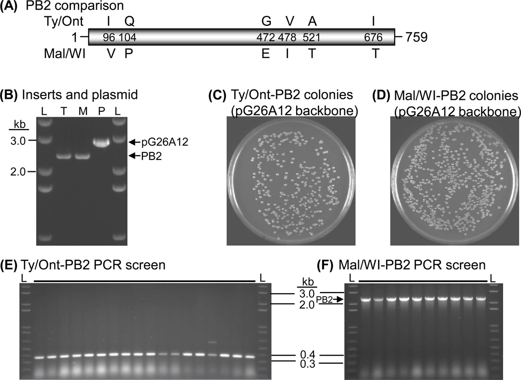
However, in the process of cloning a highly pathogenic H5N9 avian influenza A virus A/Turkey/Ontario/7732/1966 (Ty/Ont) from an M-RTPCR genomic library, no clones containing the PB2 gene segment were obtained, although all of the clones containing the other seven gene segments were acquired. PB2 specific primers were subsequently used to amplify the full-length PB2 genes of Ty/Ont, and a related H5N2 avian influenza A virus A/Mallard/Wisconsin/944/1982 (Mal/WI) as a cloning control. The sequence identity between Ty/Ont-PB2 and Mal/WI-PB2 is 91% at the nucleotide level and 99% at the amino acid level. There are only six amino acid differences between the two PB2 proteins (Fig. 1A). The Ty/Ont and Mal/WI-PB2 amplicons (~2.3 kb) were purified and analyzed by spectrometry and agarose gel electrophoresis with the StuI linearized pG26A12 reverse genetics plasmid (~2.9 kb) (Fig. 1B). Recombination-based In-Fusion® cloning reactions were set up in parallel to clone these two different PB2 genes. Briefly, 100 ng of the purified PB2 amplicon was mixed with 50 ng of the linearized pG26A12 plasmid (molar ratio ~2:1), incubated with the enzyme and buffer from the In-Fusion Advantage Kit (Clontech, Mountain View, CA) in a 10 µl reaction volume for 15 min at 37°C, followed by 15 min at 50°C. The volume was brought up to 50 µl by Tris-EDTA buffer and 3 µl was used to transform 50 µl One-Shot Top10 chemically competent cells (Invitrogen, Carlsbad, CA). The bacterial transformations were incubated at 37°C for 1 hour and a tenth of the cultures were spread onto LB agar plates containing 100 µg/ml ampicillin and incubated overnight at 37°C. The two transformation reactions generated similar size and number of colonies (Fig. 1, C and D). To avoid amplification in liquid culture, which often results in loss of unstable inserts, colonies were screened directly from the plates by PCR with primers Uni12/Inf1 (5’-GGGGGGAGCGAAAGCAGG-3’) and Uni13/Inf1 (5’-CGGGTTATTAGTAGAAACAAGG-3’), which are identical to the flanking region of the inserts (Zhou et al., 2009). More than 100 colonies from the Ty/Ont-PB2 transformation were screened, but only clones containing either very short inserts (~0.4 kb) or occasionally longer-than-expected inserts (~3.3 kb) were identified; none of the clones contained the correct length PB2 (~2.3 kb) (Fig. 1E, and data not shown). In contrast, 10 of 10 colonies screened for the Mal/WI-PB2 transformation had the correct length insert (Fig. 1F). Ultimately, more than 500 colonies from various Ty/Ont-PB2 transformations were screened, but none of them contained the correct length insert. A short insert and the longer-than-expected insert from a Ty/Ont-PB2 transformation were sequenced. The short insert resulted from a large deletion (~2 kb) in the PB2 gene, whereas the long insert resulted from a ~1 kb insertion at nucleotide position 1840 in the Ty/Ont-PB2 gene (mRNA sense). Although it is unclear how the deletion creating the small inserts occurred, the data suggest it occurred within the bacteria or it was selected from rare small fragments amplified in the RT-PCR reaction. This deletion does not appear to result from the In-Fusion cloning reaction because the short inserts were also obtained commonly in ligation based, and Topo TA based reactions using other plasmids. Interestingly, analysis of the clone containing the long insert demonstrated that it was related most closely to the bacterial IS10 transposase. Selection of this very rare transposase insertion event clearly indicates that the Ty/Ont-PB2 was under strong negative selection pressure in E. coli.
Fig. 1.
Instability of Ty/Ont-PB2 in a standard reverse genetics plasmid. (A) Diagram of the PB2 gene segment (positive sense) of Ty/Ont and Mal/WI illustrating the position of their six amino acid differences. (B) Agarose gel electrophoresis of PB2 genes of Ty/Ont (T) or Mal/WI (M) after amplification by RT-PCR, and the StuI linearized reverse genetics plasmid pG26A12 (P). L, 1kb plus DNA marker ladder (Invitrogen). (C) Colonies generated from Ty/Ont-PB2 recombination reaction. (D) Colonies generated from Mal/WI-PB2 recombination reaction. (E–F) Representative agarose gel electrophoresis of PCR amplicons obtained from colonies derived from the (E) Ty/Ont-PB2 recombination reaction (none contained an insert of correct length), or the (F) Mal/WI-PB2 recombination reaction (all contained an insert of correct length).
Aware of the selection disadvantage of the Ty/Ont-PB2 in E. coli, different bacterial stains, including Top10, DH5α, DH10B, Stbl2, Stbl3 (Invitrogen), XL1-Blue, XL10-Gold (Stratagene, La Jolla, CA), Fusion-Blue (Clontech) and a pcnB gene mutant strain for lower plasmid copy number (Lopilato et al., 1986) (kindly provided by Marlene Belfort) were transformed. Various culture media including LB, SOC, TB and 2xTY, various temperatures including 20°C, 25°C, 28°C and 37°C, and various ampicillin concentrations including 25 µg/ml, 50 µg/ml and 100 µg/ml were also attempted. Although more than 2000 colonies generated from these various strategies were screened, the correct pG26A12-Ty/Ont-PB2 construct was not obtained.
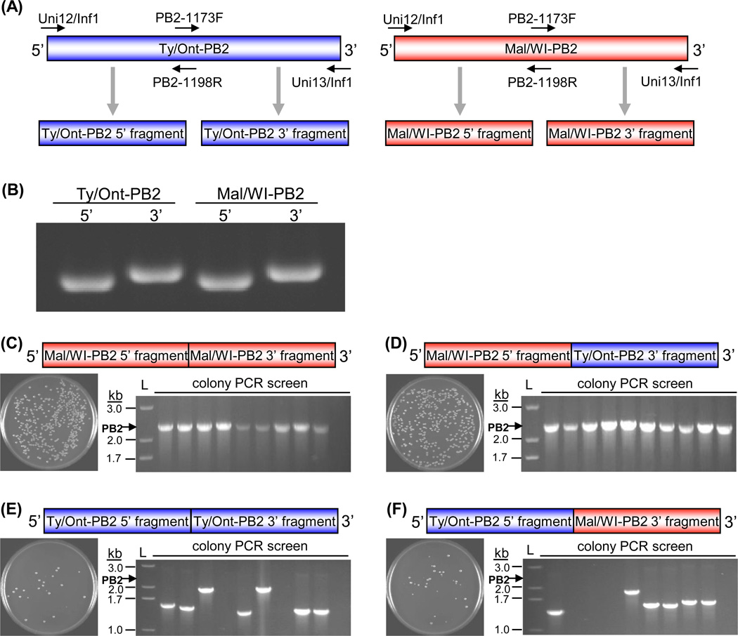
The resistance of a DNA sequence to cloning in plasmids can be caused by different factors, including long repeats, AT-rich sequences, secondary/tertiary structures, promoters, or generation of toxic proteins. In some cases, the reasons for difficulty in cloning are not well defined. To confirm that the difficulty in cloning of Ty/Ont-PB2 was sequence specific, the stability of chimeric PB2 genes from Ty/Ont and Mal/WI was analyzed. Two pairs of primers identical to both PB2 genes were designed to amplify each gene as two fragments, with overlapping regions for In-Fusion cloning (Uni12/Inf1 and PB2-1198R (5’-CGTCTCTCCCACTTACTATTAGCTG-3’) for the 5’ fragment of both PB2 genes, PB2-1173F (5’-CCAGCTAATAGTAAGTGGGAGAGACG-3’) and Uni13/Inf1 for the 3’ fragment of both PB2 genes) (Fig. 2, A and B). Different combinations of the 5’ and 3’ fragments from Ty/Ont and Mal/WI were mixed with linearized pG26A12 at a molar ratio of 2:2:1, incubated together using In-Fusion Advantage cloning kit (Clontech) and used to transform One-Shot Top10 competent cells (Invitrogen). More colonies were formed from combinations containing the 5’ fragment of Mal/WI-PB2 ((Mal/WI-5’ + Mal/WI-3’) and (Mal/WI-5’ + Ty/Ont-3’)) (Fig. 2, C and D) than from combinations containing the 5’ fragment of Ty/Ont-PB2 ((Ty/Ont-5’ + Ty/Ont-3’) and (Ty/Ont-5’ + Mal/WI-3’)) (Fig. 2, E and F). PCR across the inserts showed that almost all of the (Mal/WI-5’ + Mal/WI-3’) and (Mal/WI-5’ + Ty/Ont-3’) combinations generated clones containing PB2-length inserts (Fig. 2, C and D), while the (Ty/Ont-5’ + Ty/Ont-3’) and (Ty/Ont-5’ + Mal/WI-3’) combinations failed to generate any clones containing PB2 length inserts (Fig. 2, E and F). This data demonstrates that the 5’ fragment of Ty/Ont-PB2 is under negative selection pressure, although the fact that the IS10 transposase was inserted in the 3’ fragment (~1840bp) indicates the problem is more complicated. It is likely that exchanging smaller regions of the Ty/Ont and Mal/WI PB2 genes would identify a region within the Ty/Ont-PB2 responsible for this cloning difficulty and it could be replaced with the corresponding region of the Mal/WI-PB2. However, any modification in the PB2 gene (even silent mutations) will deviate from the goal of rescuing an authentic Ty/Ont virus.
Fig. 2.
The instability of Ty/Ont-PB2 is sequence specific. Chimeric PB2 genes were generated by PCR amplification of overlapping 5’ and 3’ fragments of Ty/Ont-PB2 and Mal/WI-PB2, which were subsequently recombined together, and with the reverse genetics plasmid (pG26A12) by In-Fusion cloning. (A) Diagram of the PCR amplification strategy used to generate 5’ and 3’ fragments of Ty/Ont-PB2 and Mal/WI-PB2. Uni12/Inf1 and PB2-1198R for amplification of 5’ fragment, PB2-1173F and Uni13/Inf1 for amplification of 3’ fragment. The 5’ and 3’ fragments have overlapping regions with each other and with the reverse genetics plasmid pG26A12 (Zhou et al., 2009). (B) Agarose gel electrophoresis of the purified PCR amplicons of the Ty/Ont-PB2 5’ and 3’ fragments and the Mal/WI-PB2 5’ and 3’ fragments used for recombination-based cloning. (C–F) Colonies formed on LB agar plates after 18 hours of incubation at 37°C and directly screened by PCR across inserts for the: (C) Mal/WI-5’ + Mal/WI-3’ combination, (D) Mal/WI-5’ + Ty/Ont-3’ combination, (E) Ty/Ont-5’ + Ty/Ont-3’ combination, or (F) Ty/Ont-5’ + Mal/WI-3’ combination. L indicates the 1kb plus DNA marker ladder (Invitrogen). The first 10 lanes of each PCR screen are shown as a representative result.
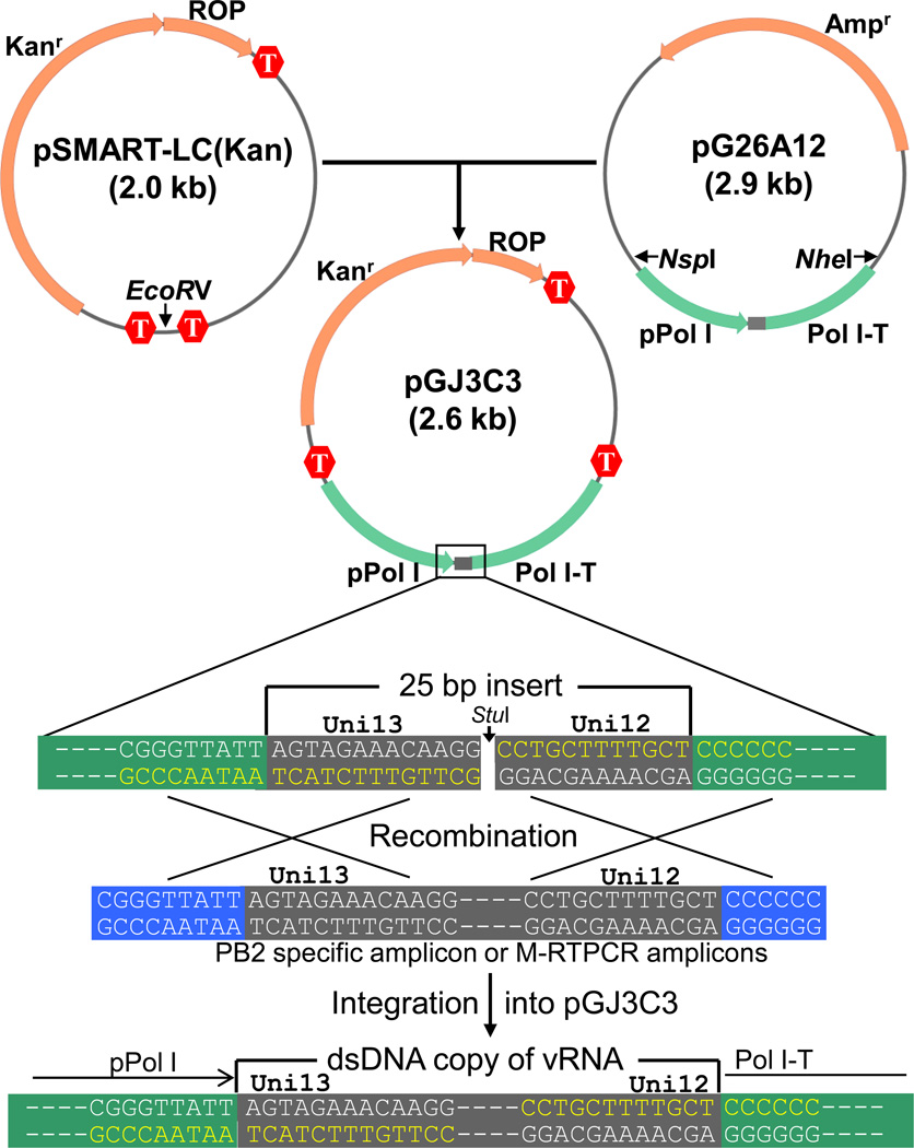
Since different bacterial strains and culture conditions did not help to clone the Ty/Ont-PB2 into the standard reverse genetics plasmid, the reverse genetics plasmid was modified to improve the stability of toxic inserts. Different commercial plasmids were tested to clone the full-length Ty/Ont-PB2, including pPCR-script (Stratagene), pcDNA3.1/V5-His-TOPO (Invitrogen) and pSMART-LC (Kan) (Lucigen, Madison, WI). Only the pSMART-LC (Kan) plasmid incorporated the correct length PB2 gene segment. The pSMART-LC(Kan) plasmid is a low copy plasmid (15~20 copies/cell) and contains transcriptional terminators at various sites to prevent transcription into or out of the inserted DNA. The successful cloning of the Ty/Ont-PB2 into pSMART-LC (Kan) suggested that the pSMART-LC (Kan) backbone may provide enhanced stability for unstable Influenza A virus genes. So the region from the Pol I promoter to the terminator was excised from the pG26A12 plasmid and ligated into the EcoRV linearized plasmid pSMART-LC (Kan) (Fig. 3). The resulting plasmid pGJ3C3 has all the features of the pSAMRT-LC vector for cloning stability as well as the Pol I promoter and terminator for influenza A vRNA generation and a Uni13/12 linker for recombination-based cloning (e.g., In-Fusion cloning) (Fig. 3). Although the 13 nucleotides at the 5’ end and the 12 nucleotides at the 3’ end of influenza vRNAs are highly conserved, a natural variation (U/C) occurs at position 4 at the 3' end. Therefore, a sister plasmid of pGJ3C3 (pGJ6B6) was also generated to clone gene segments containing the C4 variation.
Fig. 3.
Generation of recombination-based low-copy reverse genetics plasmid to clone unstable influenza A virus genes. The recombination-based reverse genetics plasmid pG26A12 (Zhou et al., 2009) has a 25 bp linker (Uni13 + Uni12, see text) between the RNA polymerase I promoter (pPol I) and terminator (Pol I-T) that was designed for In-Fusion cloning of any influenza A virus gene segment. The region containing the Pol I promoter and terminator were excised from pG26A12 by digestion with NspI and NheI, blunt-ended by mung bean nuclease digestion, and ligated into the EcoRV linearized pSMART plasmid to generate pGJ3C3 plasmid. All influenza A gene segments have identical terminal sequences to the StuI linearized plasmid and can be cloned using this recombination-based method. M-RTPCR amplicons have a few additional nucleotides (introduced by primers, shown in blue background) flanking the influenza gene segments to improve recombination efficiency. When amplifying a specific gene segment, adding these same additional nucleotides to the gene specific terminal primers also increases the efficiency of this recombination-based cloning technique. T, prokaryotic transcriptional terminator; ROP, "Repressor of Primer" copy number gene; Kanr, kanamycin resistance gene; Ampr, ampicillin resistance gene.
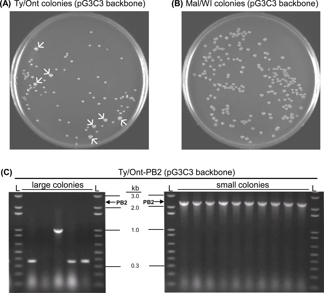
The Ty/Ont-PB2 and Mal/WI-PB2 PCR segments were then inserted into pGJ3C3 using the In-Fusion reaction, transformed into Top10 cells and incubated overnight at 37°C on LB Agar plates containing 30 µg/ml kanamycin. Except for a few large colonies, almost all of the Ty/Ont-PB2 colonies (Fig. 4A) were smaller than those generated from the Mal/WI-PB2 transformation (Fig. 4B). PCR screening of Ty/Ont-PB2 demonstrated that the large colonies contained plasmids that incorporated incorrect length inserts, whereas the small colonies contained plasmids that incorporated appropriate length inserts (Fig. 4C), which were verified by nucleotide sequencing.
Fig. 4.
Ty/Ont-PB2 was cloned successfully into the new low-copy reverse-genetics plasmid pGJ3C3. (A) The colonies derived from transformation with the Mal/WI-PB2 recombination reaction were large and uniform. (B) The majority of the colonies derived from the Ty/Ont-PB2 recombination reaction were small; however, a few large colonies (arrows) were present. (C) Agarose gel electrophoresis of amplicons from the PCR screen of colonies from the Ty/Ont-PB2 recombination reaction showed that the large colonies did not contain full-length PB2, whereas, the small colonies contained full-length PB2 inserts (2.3 kb). The length of specific fragments of the 1kb plus DNA marker ladder (L) are indicated for reference.
To demonstrate that pGJ3C3 is functional for reverse genetics, the authentic Ty/Ont virus was rescued by co-transfection of 8 plasmids expressing the Ty/Ont genomic vRNAs and 4 plasmids expressing the WSN PB2, PB1, PA and NP proteins. The influenza virus rescue process followed established methods (Hoffmann et al., 2000; Neumann et al., 1999; Zhou et al., 2010; Zhou et al., 2011), with minor modifications. Briefly, 15 µl of Lipofecatamine 2000 (Invitrogen) was used to co-transfect 0.045 µg of WSN-PA and 0.45 µg of each the other 11 plasmids into 293T cell monolayers in 35m m tissue culture treated dish, and incubated at 37°C with 5% CO2 for 8 hours. Then medium was replaced with Opti-MEM I supplemented with 2 µg/ml TPCK-trypsin (Worthington, NJ) and 2% antibiotic-antimycotic (Invitrogen). Five days post transfection, virus containing supernatants were collected and injected into the allantoic cavity of 10-day-old embryonated chicken eggs (Sunrise Farms, NY). Allantoic fluid was collected 2 days later, and the viral titer was determined to be 3 × 10⁷ TCID50/ml by TCID50 assay. All experiments involving live viruses were performed in an enhanced biosafety level 3 (BSL3+) containment laboratory approved for such use by the Centers for Disease Control and Prevention and the U.S. Department of Agriculture.
In summary, the low-copy pGJ3C3 reverse genetics plasmid can be used for cloning unstable influenza A virus gene segments. The toxic/unstable gene segment can be any of the eight segments that compose the influenza A virus genome, and this impedes tremendously the process of recombinant virus creation. Empowered by the M-RTPCR method and a recombination-based cloning strategy, the low-copy reverse genetics plasmids pGJ3C3 and pGJ6B6 can be used to clone any genomic RNA segment of influenza A viruses efficiently and will facilitate the rescue of recombinant viruses for basic research and vaccine production.
Acknowledgements
We thank Dr. Yoshihiro Kawaoka for providing the pHH21 reverse genetics plasmid, Dr. Marlene Belfort for providing the pcnB mutant bacteria strain, Rachel Hinckley, Amber Singh for experimental assistance, and the Wadsworth Center's Applied Genomic Technologies Core for nucleotide sequencing. These studies were funded primarily by NIH/NIAID R21AI 057941-02. D.E.W and B.Z. were also supported by NIH/NIAID P01AI059576-04.
Footnotes
Publisher's Disclaimer: This is a PDF file of an unedited manuscript that has been accepted for publication. As a service to our customers we are providing this early version of the manuscript. The manuscript will undergo copyediting, typesetting, and review of the resulting proof before it is published in its final citable form. Please note that during the production process errors may be discovered which could affect the content, and all legal disclaimers that apply to the journal pertain.
References
- Fodor E, Devenish L, Engelhardt OG, Palese P, Brownlee GG, Garcia-Sastre A. Rescue of influenza A virus from recombinant DNA. J. Virol. 1999;73:9679–9682. doi: 10.1128/jvi.73.11.9679-9682.1999. [DOI] [PMC free article] [PubMed] [Google Scholar]
- Hoffmann E, Neumann G, Kawaoka Y, Hobom G, Webster RG. A DNA transfection system for generation of influenza A virus from eight plasmids. Proc. Natl. Acad. Sci. 2000;97:6108–6113. doi: 10.1073/pnas.100133697. [DOI] [PMC free article] [PubMed] [Google Scholar]
- Lopilato J, Bortner S, Beckwith J. Mutations in a new chromosomal gene of Escherichia coli K-12, pcnB, reduce plasmid copy number of pBR322 and its derivatives. Mol. Gen. Genet. 1986;205:285–290. doi: 10.1007/BF00430440. [DOI] [PubMed] [Google Scholar]
- Neumann G, Watanabe T, Ito H, Watanabe S, Goto H, Gao P, Hughes M, Perez DR, Donis R, Hoffmann E, Hobom G, Kawaoka Y. Generation of influenza A viruses entirely from cloned cDNAs. Proc. Natl. Acad. Sci. 1999;96:9345–9350. doi: 10.1073/pnas.96.16.9345. [DOI] [PMC free article] [PubMed] [Google Scholar]
- Palese P, Shaw ML. Orthomyxoviridae: the viruses and their replication. In: Knipe DM, Howley PM, Griffin DE, Lamb RA, Martin MA, Roizman B, Straus SE, editors. Fields Virology. Philadelphia, PA: Lippincott Williams and Wilkins; 2007. pp. 1647–1690. [Google Scholar]
- Wright PF, Neumann G, Kawaoka Y. Orthomyxoviruses. In: Knipe DM, Howley PM, Griffin DE, Lamb RA, Martin MA, Roizman B, Straus SE, editors. Fields Virology. Philadelphia, PA: Lippincott Williams and Wilkins; 2007. pp. 1691–1740. [Google Scholar]
- Zhou B, Donnelly ME, Scholes DT, George K, Hatta M, Kawaoka Y, Wentworth DE. Single-reaction genomic amplification accelerates sequencing and vaccine production for classical and swine origin human influenza A viruses. J. Virol. 2009;83:10309–10313. doi: 10.1128/JVI.01109-09. [DOI] [PMC free article] [PubMed] [Google Scholar]
- Zhou B, Li Y, Belser JA, Pearce MB, Schmolke M, Subba AX, Shi Z, Zaki SR, Blau DM, Garcia-Sastre A, Tumpey TM, Wentworth DE. NS-based live attenuated H1N1 pandemic vaccines protect mice and ferrets. Vaccine. 2010;28:8015–8125. doi: 10.1016/j.vaccine.2010.08.106. [DOI] [PMC free article] [PubMed] [Google Scholar]
- Zhou B, Li Y, Halpin R, Hine E, Spiro DJ, Wentworth DE. PB2 residue 158 is a pathogenic determinant of pandemic-H1N1 and H5 Influenza A viruses in mice. J. Virol. 2011;85:357–365. doi: 10.1128/JVI.01694-10. [DOI] [PMC free article] [PubMed] [Google Scholar]


