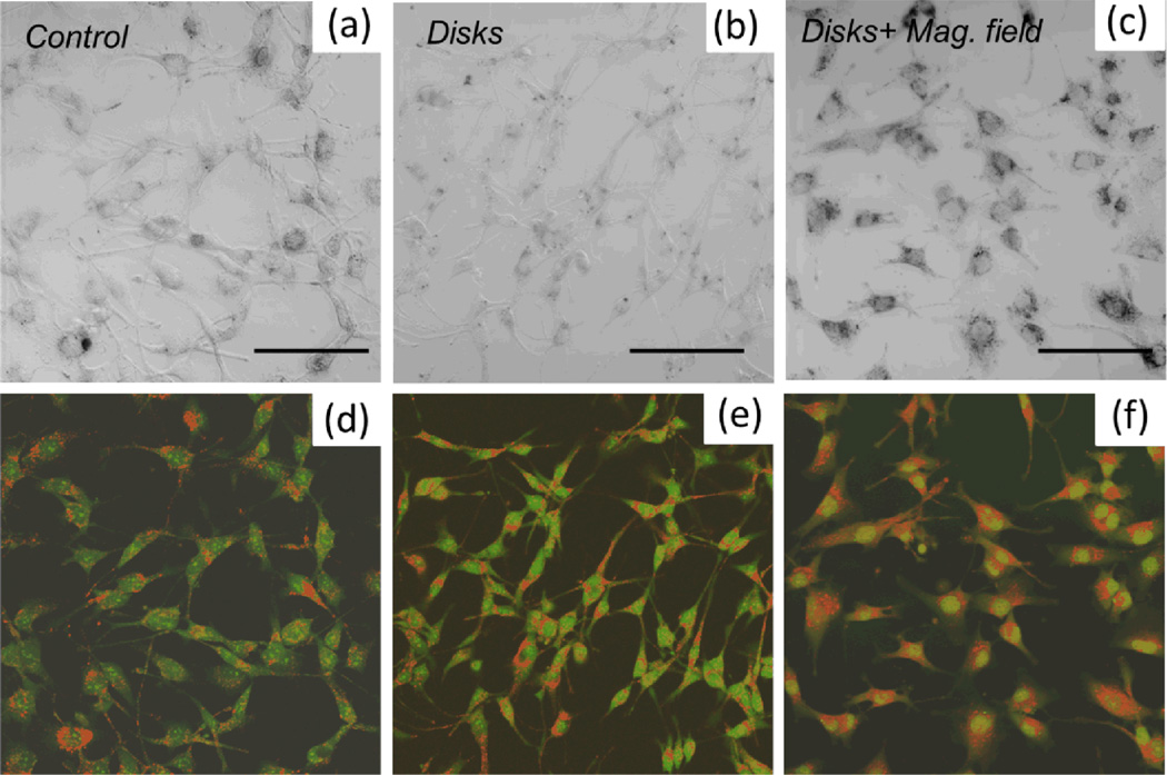
Figure 3.
DIC image of (a) control human glioblastoma cells A172, (b) cells cultured with ferromagnetic disks, (c) cells cultured with magnetic disks and subjected to a.c. magnetic field. Scale bar 100 µm, 20x magnification objective. Each sample was stained with acridine orange/ethidium bromide for assessing the apoptotic changes. Panels (d–f) show the corresponding confocal fluorescence images. Only the cells which were exposed to a.c. magnetic field (panel (f)) exhibit the signs of early apoptosis as indicated by enhanced nuclear staining. At the same time, by comparing the distribution of AO/EB staining in panels (d) and (e), one can conclude that culturing glioblastoma cells with magnetic disks present of their membrane does not compromise the cell viability.

