


Figure 2.
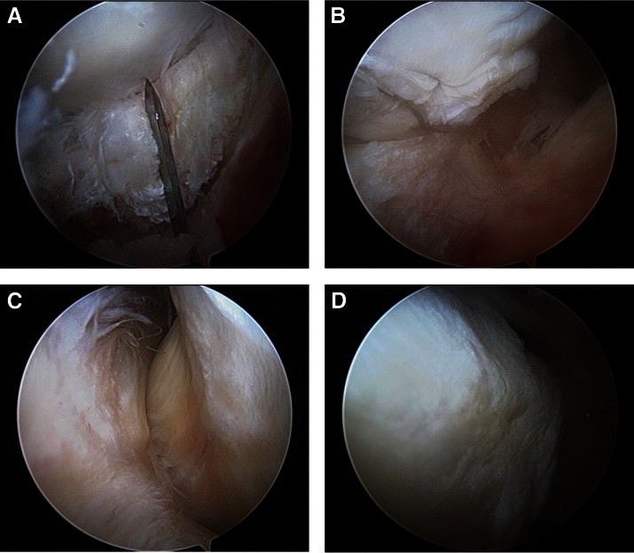
Expansile view of peripheral compartment with arthroscope in anterior portal, viewing distally and laterally. (A) Capsulotomy with arthroscopic scalpel through iliofemoral ligament between gluteus minimus and iliocapsularis muscles. (B) T capsulotomy down anterior aspect of femoral neck to intertrochanteric line. (C) View of lateral synovial folds. (D) View of medial synovial folds.