
Figure 4.
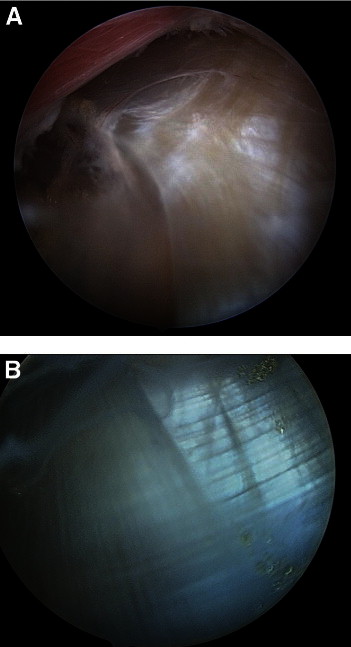
(A) Proximal-to-distal view of falciform ligament in a right shoulder, with the arthroscope through the accessory anterolateral portal, viewing distal within the subdeltoid bursae. The intersection between the longitudinal fibers of the biceps sheath and the transverse fibers of the superior edge of the pectoralis major should be noted. (B) Close-up view of intersecting fibers that make up falciform ligament. The LHB lies just beneath this visual landmark.