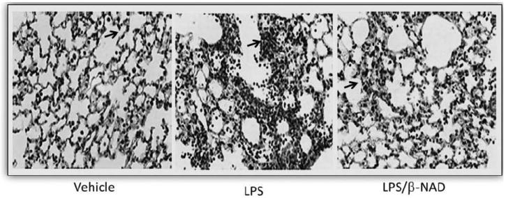
FIGURE 3.

Histopathology. β-NAD inhibits the inflammation in lungs of mice in LPS-induced ALI. Lungs perfused free of blood, were immersed in 4% buffered paraformaldehyde at 4°C for 18 hours prior to histological evaluation by hematoxylin and eosin (H&E) staining. H&E staining was done by deparafinizing and hydrating the slides to water. The slides were stained in Harris hematoxylin for 15 minutes and eosin for 30 seconds. The slides were dehydrated, cleared, and mounted with cytoseal. Histological analysis of the lung tissue obtained from the control mice exposed to PBS showed minimal infiltration of neutrophils. In contrast, mice exposed to LPS for 24 hours produced prominent neutrophil infiltration and that was attenuated in LPS/β-NAD simultaneously.
