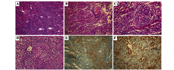
Figure 1.
Pathological characteristics of pharyngeal FDCS. (A) Tumor cells arranged in a diffusing sheet-like distribution, partially in the storiform pattern (case 2). (B) Tumor cells arranged in the whorl pattern (case 3). (C) Indistinct cell borders and slightly eosinophil-stained cytoplasm of tumor cells. Infiltrating small lymphocytes and tissue cells were observed in the background (case 3). (D) Nuclei of tumor cells were irregular, round- or spindle-shaped, containing delicate chromatin and small nucleoli. Clear mitotic counts were observed (case 1). (E) Tumor cells were positive for podoplanin (case 3). (F) Tumor cells were negative for LCA, but the lymphocytes scattered in the background were positive for LCA (case 3). FDCS, follicular dendritic cell sarcoma; LCA, leukocyte common antigen.

