
Figure 6.
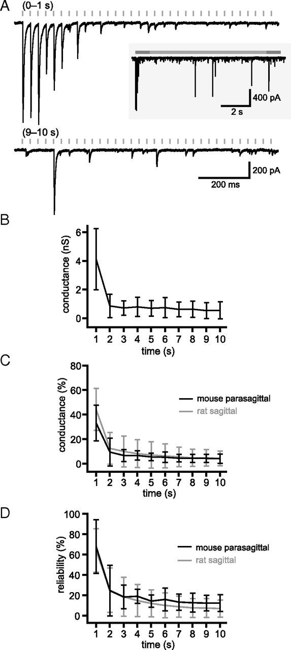
GPe–STN synaptic depression is not the result of transection of GPe–STN axons. A, Example of unitary synaptic transmission after stimulation of the GPe in a mouse parasagittal brain slice that preserves GPe–STN connectivity. The first and 10th seconds of stimulation at 33 Hz are shown. Inset, The entire stimulation period. B–D, Population data (n = 5). Mean conductance (B), mean normalized conductance (C), and reliability of transmission (D) against time. C, D, Gray traces represent overlay of unitary rat GPe–STN data for comparison.