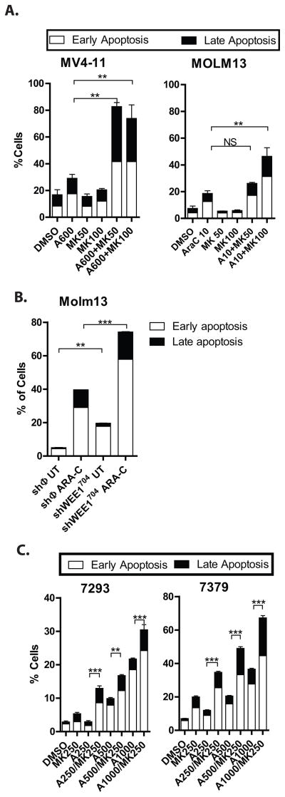
Figure 7. Inhibition of WEE1 in combination with cytarabine enhances the apoptotic effects of cytarabine in AML cell lines and primary AML cells.
A. MK1775 plus cytarabine results in more apoptosis than cytarabine alone in AML cell lines. Cells treated as in 6A were stained for annexin V and with 7-AAD and analyzed by flow cytometry. The percentage of early apoptotic (white bar) and late (apoptotic) cells are shown. B. Knockdown of WEE1 sensitizes AML cells to apoptosis induced by cytarabine. Molm13 cells transduced with a non-silencing control vector (shφ) or an shRNA directed against WEE1 (as in Figure 5D) were treated with cytarabine at 25nM for 72 hours. The percentage of apoptotic cells was assessed as in Figure 7A. C. MK1775 plus cytarabine results in more apoptosis than cytarabine alone in primary AML cells. Primary AML cells were cultured with cytarabine and/or MK1775 at the indicated concentrations for 72 hours and the percentage of apoptotic cells was assessed as in Figure 7A. Asterisks indicate statistical significance (ANOVA).

