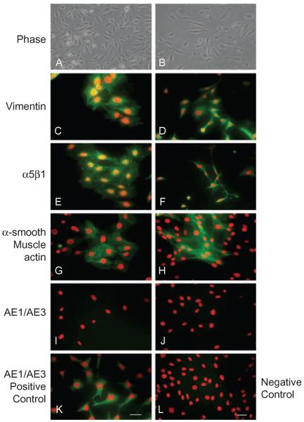
FIGURE 2.
Characterization of immortalized WT and 7ko fibroblasts. WT and 7ko fibroblasts were visualized by phase contrast [WT in (A), 7ko in (B)] and immunostained for vimentin [WT in (C), 7ko in (D)], integrin α5β1 [WT in (E), 7ko in (F)], and epithelial keratin [using AE1/AE3 antibody; WT in (I), 7ko in (J)]. A subset of cells was stimulated with TGF-β and stained for α-SMA (myofibroblast marker) [WT in (G), 7ko in (H)]. Corneal epithelial cells immunostained for epithelial keratin served as a positive control (K), and TGF-β-stimulated corneal epithelial cells immunostained for α-SMA served as a negative control (L). Scale bar, 50 μm.

